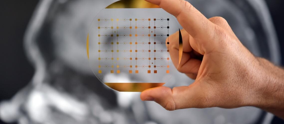
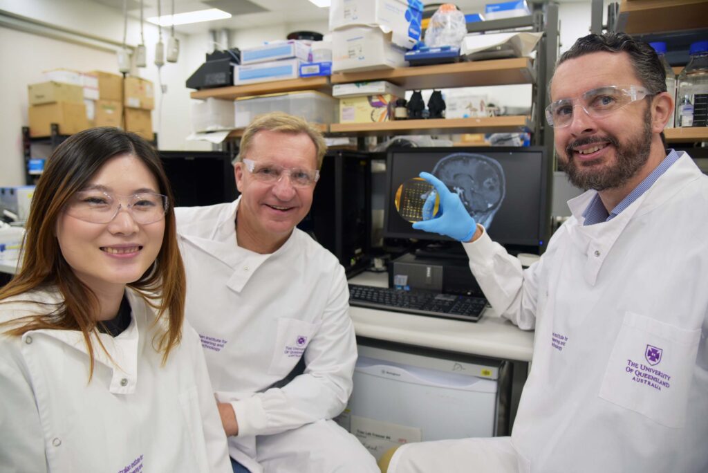
Australian researchers at the University of Queensland's Australian Institute for Bioengineering and Nanotechnology (AIBN) have developed a nanoscale diagnostic chip that may revolutionise how doctors assess the effectiveness of treatment of brain cancer patients by providing the first non-invasive method for tracking glioblastoma progress using a simple blood sample. The Phenotype Analyser Chip is able to identify micro-scale cellular bundles (vesicles) that contain molecular information from brain tumours, which are released into the bloodstream and can cross the blood-brain barrier.
Physicians currently face a significant limitation when treating glioblastoma, the most aggressive type of brain cancer. Physicians must either perform an invasive biopsy or wait until the tumour grows sufficiently to be detected by MRI, thereby allowing them to evaluate whether the treatment is working before making treatment adjustments. The new chip-based technology may provide physicians with real-time data on the tumour’s response to treatment, potentially accelerating the development of more effective treatments for glioblastoma, which has seen little therapeutic progress.
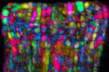
The engineering breakthrough lies in detecting the extracellular vesicles (vesicles), tiny membrane-bound structures that cells release into the bloodstream. Vesicles are approximately 30-150 nm in diameter and serve as cellular messengers, containing proteins, genetic material and other molecular components that reflect the original cell of origin. When brain tumour cells release these vesicles,s they must cross the blood-brain barrier. This selectively permeable membrane usually prevents most substances from crossing between the bloodstream and brain tissue.
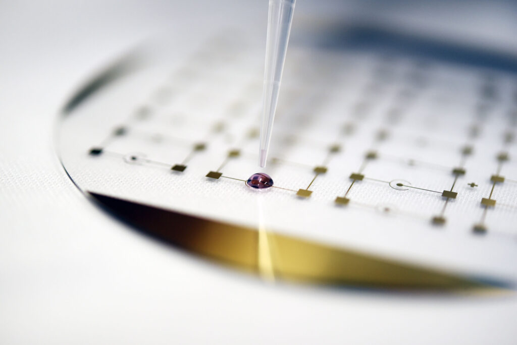
The PhenotypeAnalysser Chip employs nanotechnology to capture and analyse minute particles in blood samples. The device contains engineered surfaces with molecular recognition elements that will selectively bind to vesicles originating from glioblastoma cells. After binding to the engineered surface, the chip enables examination of the vesicle’s molecular content, providing information about the tumour’s current status and biological activity.
This methodology addresses a long-standing engineering problem in neuro-oncological diagnostics. Although the blood-brain barrier protects brain tissue from many harmful substances, it limits the number of tumour-derived biomarkers that reach detectable levels in blood samples. However, due to the unique membrane properties of extracellular vesicles, they can cross the blood-brain barrier with their molecular cargo intact.
How the Phenotype Analyzer Chip Works
Innovative Engineering Approach
The Phenotype Analyser Chip is built on the integration of three technologies: nanotechnology, microfluidics, and molecular engineering.
Process Overview
At the heart of the Phenotype Analyser Chip is a multistage analytical process that commences once blood enters the microscopic channels etched into the chip's surface.
Detection Mechanism
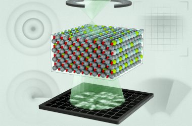
Engineers have created nanostructures at the chip's surface using lithography (a technique also used in semiconductor manufacturing) to achieve a high surface-area-to-volume ratio. The nanostructures were then chemically modified with capture molecules (antibodies), which are capable of recognising proteins on the surface of glioblastoma-derived vesicles.
Capture Process
The interaction between the blood and the nanostructures occurs within microchannel networks. The fluid dynamics of the microchannel network were optimised by engineers to maximise interaction between the vesicles and the capture molecules, while maintaining adequate flow rates for practical clinical application. Factors including the geometry of the microchannels, the flow rate of the blood, and the chemical properties of the surface were all optimised to provide selective capture efficiencies greater than 70% for vesicles derived from glioblastomas.
Analysis
Once vesicles have bound to the surface of the nanostructures, the Phenotype Analyzer Chip uses a variety of techniques to analyse the vesicle content. Optical techniques, such as surface plasmon resonance or fluorescence microscopy, may be employed to detect specific protein markers present on the vesicle surface. In addition, the PhenotypeAnalyserr Chip may include electrical detection mechanisms that measure changes in electrode impedance or conductance resulting from vesicle binding to the electrode surface.
Engineering Challenge
While detection of vesicle binding is a significant achievement, it is not the only engineering challenge associated with the Phenotype Analyzer Chip. Specifically, glioblastoma-derived vesicles are extremely rare in blood samples, possibly representing as few as one in a million total vesicles. To address this issue, engineers employed a hierarchical capture strategy. Initially, a series of capture zones was employed to remove vesicles from non-tumour sources, and then subsequent zones employed increasingly specific molecular recognition elements to isolate brain tumour-derived particles.
Thermal Management
Another engineering consideration was the temperature control of the Phenotype Analyser Chip. Vesicles remain stable at body temperature, but will rapidly degrade under altered conditions. Therefore, the chip design incorporated thermal management capabilities to maintain optimal temperatures during sample processing, thereby ensuring vesicle stability throughout the analysis.
Multiplexed Analysis
One final advantage of the microfluidic architecture of the Phenotype Analyser Chip is its ability to perform multiplexed analysis. Multiplexed analysis involves simultaneous testing for multiple biomarkers from a single sample. Engineers achieved this capability by developing a parallel-channel architecture in which each region of the chip contained distinct capture molecules. This parallel processing approach increased diagnostic confidence by confirming tumour presence based on multiple independent measurements.
Validation studies with over 40 brain cancer patients demonstrated that this chip can distinguish between various stages of glioblastoma progression. Moreover, as previously stated, this technology has shown great potential for detecting early treatment responses, which may occur many weeks before such responses are observed with conventional imaging methods.
Although glioblastoma detection is an immediate application, the engineers developing this technology envision broader applications. The principle behind the detection of brain-tumour vesicles can be applied to the monitoring of other neurological disorders in which characteristic extracellular vesicles are released by damaged cells across the blood-brain barrier.

In previous work from this research group, researchers have found that traumatic brain injuries (TBI) lead to the release of brain-derived biomarkers into blood; therefore, it appears possible that this chip may aid in assessing the degree of neuroinflammation following TBI, and thus provide an objective measure for assessment of TBI, which relies almost entirely upon subjective reports of symptoms at present.
Additionally, this technology may allow for earlier diagnosis and monitoring of neurodegenerative diseases such as Alzheimer’s and Parkinson’s. Both diseases are characterised by progressive cellular damage, which is likely to produce distinct vesicle signatures long before clinical symptoms become apparent. Therefore, if the chip can detect these molecular patterns, it could enable earlier therapeutic interventions at stages in the disease process when treatments may be more effective.
Additional target areas include motor neurone disease and depression, both of which have no objective biomarkers for quantitatively measuring disease progression or treatment response. A chip-based diagnostic system could provide quantitative data to assist clinicians in making therapy decisions and expedite drug development.
However, several engineering challenges remain to be overcome to transition this technology from laboratory validation to clinical implementation. First, manufacturers must develop manufacturing processes that yield consistent chip performance across all production batches. Additionally, sample preparation protocols must be standardised to minimise variability between samples prepared in different clinical settings. Furthermore, the integration of the chip-detection system with existing laboratory equipment and workflows must be carefully considered with respect to practical constraints, including processing time and the need for trained personnel.
Finally, cost-effectiveness analysis will ultimately determine whether this technology can be widely adopted. Although the chips themselves may be produced in large quantities using established semiconductor manufacturing techniques, the requirement for highly specialised detection equipment and trained personnel to operate it may limit the initial deployment of this technology to major medical centres.
At this point in time, the development team is focused on initiating clinical trials to demonstrate the diagnostic accuracy of the chip across larger numbers of patients. In addition to demonstrating the chip’s detection capability, these studies must also assess how chip-based monitoring affects clinical decision-making and patient outcomes. If successful, this technology could provide clinicians with unprecedented real-time insights into brain tumour biology. It could potentially improve patient outcomes in individuals diagnosed with one of the most difficult-to-treat forms of cancer.
TLDR
- Australian scientists created a nanoscale chip that detects brain cancer through blood samples, eliminating the need for invasive biopsies
- The Phenotype Analyzer Chip captures microscopic cellular packages (extracellular vesicles) that cross the blood-brain barrier from glioblastoma tumours
- Technology provides real-time treatment monitoring, potentially detecting therapy responses weeks before MRI scans show changes
- Chip uses engineered nanosurfaces and microfluidics to capture tumour-derived vesicles from blood samples selectively
- Validated in 40+ brain cancer patients, showingthe ability to distinguish different stages of glioblastoma progression
- Applications could expand to Alzheimer’s, Parkinson’s, traumatic brain injury, and other neurological conditions
- Technology addresses critical gap in glioblastoma treatment monitoring for world’s most aggressive brain cancer
- Could accelerate drug development and improve patient outcomes through earlier treatment response detection