
Aerospace, Defence & MarineBreakthroughs
Organ chip space medicine technology flew on Artemis II, testing personalised bone marrow chips to study deep space radiation effects on astronaut health.
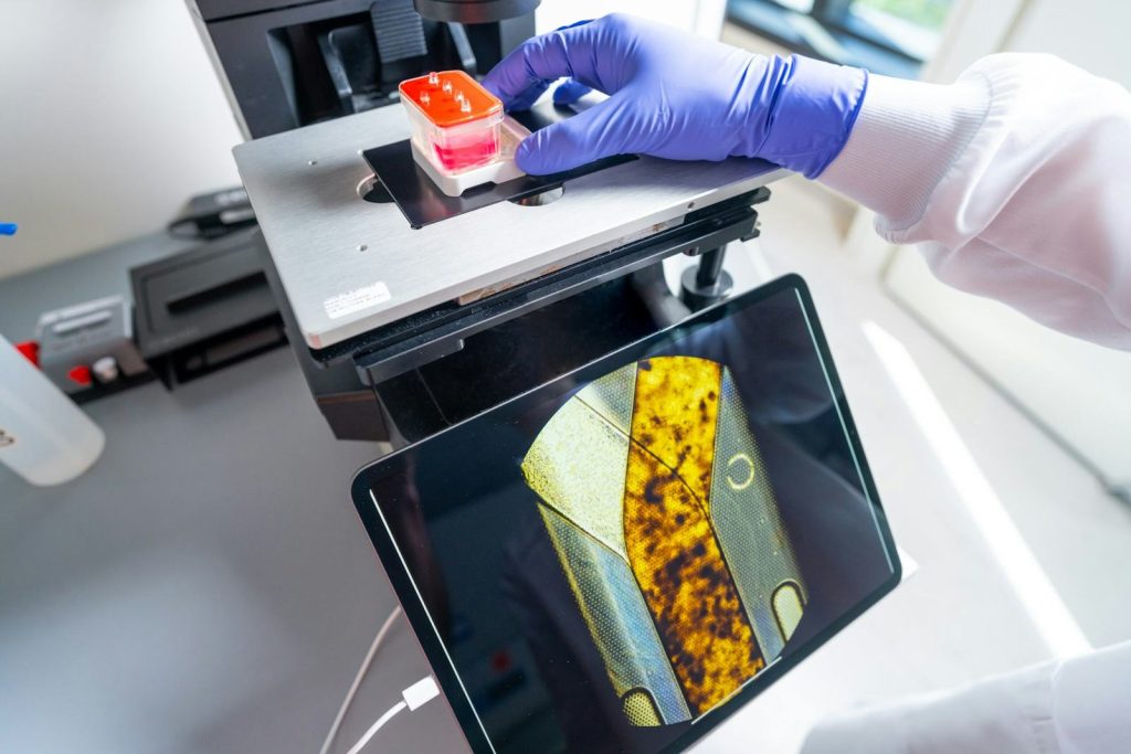
Somewhere inside the Orion spacecraft, as it swung around the far side of the Moon in early April 2026, a set of devices no bigger than USB thumb drives was quietly doing science. Each one contained living bone marrow cells, grown from blood samples taken from the four Artemis II astronauts, and maintained in fluid that mimicked the conditions inside a human body. These organ chips, developed by the Wyss Institute for Biologically Inspired Engineering at Harvard University, were measuring what happens to human tissue when it leaves the protection of Earth’s magnetic field and enters the radiation environment of deep space, as announced by the Wyss Institute.
The experiment is called AVATAR, which stands for A Virtual Astronaut Tissue AAnalogueResponse. The idea is straightforward, even if the engineering is not: rather than waiting for astronauts to return from a mission and then checking whether their bodies have changed, you send their cells along for the ride and watch what happens in real time. Bone marrow was chosen because it is particularly vulnerable to radiation. It is also the source of circulating blood cells, which show measurable alterations in some astronauts after spaceflight. By comparing the organ chips aboard Orion with a matched set kept on Earth under otherwise identical conditions, researchers can isolate the effects of radiation and microgravity from everything else.
Dr Donald Ingber, the principal investigator and founding director of the Wyss Institute, has spent more than a decade developing organ chip space medicine technology. Dr David Chou leads the research teams at both the Wyss Institute and Emulate, Inc., the company that commercialised the chips. The AVATAR experiment represents the first time personalised organ chips, containing cells from the actual crew members, have flown beyond low Earth orbit.
Previous studies of spaceflight’s effects on human health have relied on blood samples, questionnaires, and medical imaging before and after missions. These methods tell you what changed but not how or why. Organ chips offer a window into the mechanism. They contain tiny hollow channels lined with living human cells arranged to replicate the architecture and function of a specific organ. Fluid flows through the channels, mimicking blood circulation. Sensors embedded in the chip can continuously track cell behaviour, gene expression, and inflammatory markers.
For Artemis II, the bone marrow chips were seeded with haematopoietic stem cells, the precursors that produce red blood cells, white blood cells, and platelets. In deep space, outside the Van Allen belts that shield low Earth orbit from the worst of the Sun’s particle radiation, these cells face a cocktail of galactic cosmic rays and solar energetic particles that ground-based laboratories cannot fully replicate. If the chips show measurable changes in blood cell production, DNA damage rates, or inflammatory signalling, it would provide the strongest evidence yet that deep space radiation affects the blood-forming system in ways that short-duration missions in low Earth orbit do not.
The technology has implications well beyond spaceflight. If organ chips can reliably model how human tissue responds to radiation in real time, they could replace some animal testing in pharmaceutical development and cancer research, where understanding radiation effects on bone marrow is directly relevant to patients undergoing radiotherapy.
Engineering deep dive
An organ chip is a microfluidic device that combines microfabrication techniques from the semiconductor industry with cell biology. The chip itself is typically made of polydimethylsiloxane (PDMS), a flexible, transparent silicone polymer, and is bonded to a glass slide. Inside are two or more parallel channels separated by a thin, porous membrane. Living human cells are cultured on either side of this membrane, creating a tissue-tissue interface that mimics the structure of a real organ.
For a bone marrow chip, one channel is lined with endothelial cells, the cells that form blood vessel walls. At the same time, the other contains haematopoietic stem and progenitor cells embedded in a gel matrix that replicates the extracellular environment of bone marrow. A syringe pump pushes cell culture medium through the channels at controlled flow rates, simulating blood circulation. The flow rate, typically a few microlitres per hour, is calibrated to produce shear stresses similar to those experienced by cells in living capillaries. If you have studied fluid dynamics in A-level Physics, you will recognise this as an application of Poiseuille's law, which describes the relationship between flow rate, channel dimensions, fluid viscosity, and pressure drop.
The porous membrane between the channels serves a dual purpose. It provides structural support for the cells while allowing molecules, nutrients, waste products, and signalling factors to pass between the two tissue layers. The pore size, typically between 0.4 and 8 micrometres, is selected to permit molecular diffusion while preventing cell migration between compartments. This selective permeability is analogous to the basement membranes that separate tissues in the body, a concept covered in A-level Biology.
Fabricating these chips at the scale needed for spaceflight required solving several practical engineering problems. The devices must survive launch vibration, operate in microgravity where fluid behaviour changes (surface tension dominates over gravity, so bubbles become trapped more easily), and run autonomously for the duration of the mission without a lab technician to adjust flow rates or replenish media. The Emulate team designed self-contained cartridges with pre-loaded media reservoirs and battery-powered micropumps. The entire assembly was tested on parabolic flights and the International Space Station before being cleared for the Artemis mission.
Sensors integrated into the chip measure several parameters in real time. Electrical impedance across the membrane tracks barrier function, the integrity of the cell layer. Optical sensors can detect fluorescent markers that indicate specific biological processes such as apoptosis (programmed cell death) or DNA double-strand breaks. Temperature and pH sensors ensure the culture environment remains stable. The data is logged onboard and downloaded after the mission for comparison with the Earth-based control chips.
One of the most interesting engineering aspects is personalisation. Each astronaut's organ chips were created from their own induced pluripotent stem cells (iPSCs), which are adult cells reprogrammed to behave like embryonic stem cells. These iPSCs were then differentiated into the specific cell types needed for the bone marrow chip. This means the experiment captures individual biological variation, not just an average response, opening the door to personalised risk assessment for future crews.
Links to the curriculum: A level, GCSE
The Artemis II astronauts returned to Earth on 10 April 2026. Analysis of the organ chips will take months. If the results confirm that personalised tissue models can reliably detect radiation-induced changes during a 10-day mission, the next step would be sending larger numbers of chips on longer missions, potentially including Artemis III’s planned lunar surface expedition. The ultimate goal is a personalised medical kit that travels with each astronaut, monitoring their biology as they go. We are not there yet. But the chips are back from the Moon.
Credit via wyss.harvard.edu
Aerospace, Defence & MarineBreakthroughs


Aerospace, Defence & Marine