Engineers at MIT have revealed a stunning new 3-D imaging technique which reveals how neurons connect throughout the brain.

The researchers released this image of the entire fruit fly brain.
The new method allows researches to perform large-scale, 3D imaging of brain tissue with unprecedented resolution and speed. The results are not just beautiful but can be used to locate individual neurons, trace connections between them, and visualize organelles inside neurons, over huge volumes of brain tissue
Stunning Images of how brain cells interact
The team released a series of stunning images alongside their research showing how cells within the brains of mice and fruit flies connect.

Color-coded by 3D depth – Dopaminergic neurons in the ellipsoid body of a fruit fly brain

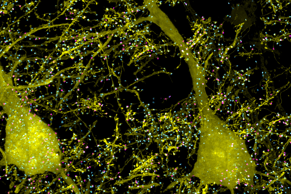
A subset of pyramidal neurons (yellow), and pairs of presynaptic (cyan) and postsynaptic (magenta) proteins associated with the neurons in the mouse primary somatosensory cortex.

Individually traced dopaminergic neurons in the right hemisphere of a fruit fly brain, innervating the fan-shaped body (green), ellipsoid body (magenta), and noduli (green).

A subset of pyramidal neurons (orange) in the mouse primary somatosensory cortex. The dendritic spines associated with the postsynaptic protein Homer1 are highlighted in yellow.
How Cortical column and whole-brain imaging works
The technique works by combining an existing method for expanding brain tissue (which means higher resolution images) with lattice light-sheet microscopy – a rapid 3-D microscopy technique.
Because the team expanded brain tissue before imaging they are able to image the tissue at a resolution of about 60 nanometers. Until this technique was developed, this type of imaging had to be done with incredibly expensive high-resolution microscopes, known as super-resolution microscopes.
The combination of these methods meant researchers could image an entire fruit fly brain, as well as large sections of the mouse brain, much faster than has previously been possible.
Alongside the challenges of marrying the two techniques researchers also had cope with the massive amounts of data generated. Each sample would create terabytes of imaging data. To process the data into a manageable form the team used a highly parallelized computational image-processing techniques to break down the data into smaller chunks, analyze it, and stitch it back together into a coherent whole.
While the technique is a breakthrough for researchers looking to map large-scale circuits within the brain it could also allow researchers in other fields to look at how cells interact. For example, seeing how HIV is able to evade the human immune system or study how cancerous cells interact with their surroundings.
In future the team want to expand the technique to trace circuits that control memory formation and recall, to study how sensory input leads to a specific behaviour, or to analyze how emotions are coupled to decision-making.
Published as Cortical column and whole-brain imaging with molecular contrast and nanoscale resolution in Science Vol 363, Issue 6424 18 January 2019
The research team included researchers from MIT, the University of California at Berkeley, the Howard Hughes Medical Institute, and Harvard Medical School/Boston Children’s Hospital


