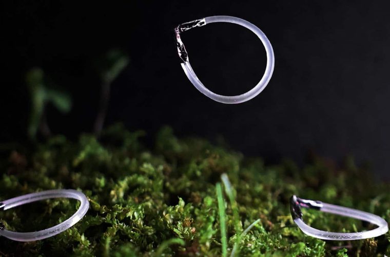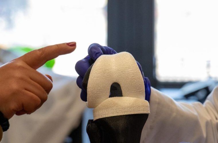
Researchers at ETH Zurich have developed a 3D bioprinted interface that replicates the biological connection between muscle and bone, addressing a fundamental challenge in biohybrid robotics and medical implant design. The system, developed at ETH Zurich’s Soft Robotics Lab under the leadership of Professor Robert Katzschmann, demonstrates stable force transmission between living tissue and synthetic materials through engineered tendon-like structures.
The biohybrid actuator addresses the persistent problem of energy loss at interfaces between soft biological tissues and rigid synthetic components. Traditional approaches to combining these materials have struggled with inefficient force transmission, limiting the practical applications of biohybrid systems in both robotics and medicine.
Working with researchers from the Institute for Bioengineering of Catalonia (IBEC) and the University of Barcelona, the ETH Zurich team focused on replicating the myotendinous junction. In this natural transition zone, tendons connect to muscle tissue. This biological structure serves as an intermediate layer with graduated stiffness properties, enabling efficient mechanical coupling between materials with different elastic moduli.
“Our solution is based on a 3D bioprinted actuator that structurally and functionally mimics the natural connection between muscle and bone,” said Miriam Filippi, lead author and researcher at the Soft Robotics Lab. The team’s approach involved printing living muscle cells alongside tendon-like anchors containing connective tissue cells onto a substrate platform.
The research team employed computer-assisted analysis to optimise the geometry and internal structure of the bioprinted actuator. The design process focused on creating a gradient of mechanical properties that would minimise stress concentrations at material interfaces while maintaining structural integrity during repeated contraction cycles.
The bioprinting process deposited multiple cell types in precise spatial arrangements, with muscle cells forming the contractile element and connective tissue cells creating the transitional tendon structure. The printed tendon component exhibits intermediate stiffness values between the compliant muscle tissue and the rigid synthetic bone analogue, facilitating mechanical impedance matching across the interface.
Initial testing demonstrated that the 3D bioprinted actuators maintained reliable contraction capabilities over extended periods of time. The stability of these contractions suggests that the engineered tissue interface can withstand the mechanical stresses associated with repeated actuation cycles without degradation of the biological-synthetic junction.
The mechanical challenge of coupling biological and synthetic materials stems from their vastly different elastic moduli. Living muscle tissue typically exhibits elastic moduli in the range of 10-100 kPa, while synthetic substrates used in robotics applications often exceed 1 GPa. This three-to four-order-of-magnitude difference creates significant stress concentrations at direct interfaces, leading to delamination, tearing, or inefficient force transfer.
The myotendinous junction in natural systems solves this problem through a hierarchical structure that gradually transitions mechanical properties. At the molecular level, collagen fibres from tendons interdigitate with muscle cell membranes through specialised protein complexes. These connections distribute mechanical loads across multiple length scales, thereby preventing localised stress concentrations that could cause failure.
In bioprinting applications, recreating this complexity requires precise control over several parameters:
- Cell density gradients: The concentration of muscle cells decreases while connective tissue cells increase along the transition zone
- Extracellular matrix composition: The ratio of collagen types I and III varies spatially to modulate stiffness
- Fibre alignment: Anisotropic organisation of structural proteins directs force transmission pathways
- Crosslinking density: Chemical crosslinks between collagen molecules determine the mechanical properties of the printed tissue
The bioprinting process itself presents unique engineering constraints. Shear stresses during extrusion must remain below thresholds that would damage cells (typically <5 kPa), while maintaining sufficient resolution to create functionally graded structures. Print speeds, nozzle diameters, and bioink rheology all influence cell viability and final tissue architecture.
Force transmission efficiency in the biohybrid system depends on several mechanisms operating simultaneously. Mechanical interlocking occurs as the printed tissue integrates with surface features on the synthetic substrate. Chemical bonding through cell-secreted adhesion proteins creates molecular-level connections. Additionally, the viscoelastic properties of the biological component provide damping that protects the interface during dynamic loading.
The computer-assisted optimisation employed by the ETH team likely involved finite element modelling to predict stress distributions under various loading conditions. Parameters such as the length and taper angle of the tendon region, the distribution of cell types, and the surface topology of the synthetic anchor points would be iteratively refined to minimise peak stresses while maximising force transmission efficiency.
Maintaining tissue viability in biohybrid systems requires careful consideration of nutrient transport. The acellular synthetic components must incorporate channels or porous structures that allow diffusion of oxygen and nutrients to the living tissue. For actuators thicker than 200 micrometres, passive diffusion becomes insufficient, necessitating perfusion systems or the incorporation of vascular networks.
Temperature regulation presents another engineering challenge. Muscle tissue generates heat during contraction, which must be dissipated to prevent thermal damage. The thermal conductivity mismatch between biological and synthetic materials can create localised hot spots that compromise tissue viability. Design strategies might include heat sinks integrated into the synthetic components or active cooling systems for high-frequency applications.
The ETH Zurich team’s testing protocols evaluated both immediate functionality and long-term stability of the biohybrid actuators. Contraction force measurements demonstrated that the engineered muscle-tendon units could generate consistent mechanical output over multiple activation cycles. The preservation of contractile function over extended culture periods indicates successful integration between the biological and synthetic components.
If these initial results translate to scaled applications, the technology could enable biohybrid systems with performance characteristics approaching those of natural musculoskeletal structures. The stability of the interface under repeated loading cycles suggests potential durability for practical applications. However, long-term studies under varied environmental conditions would be necessary to confirm clinical or industrial viability.
The immediate applications for this technology span both medical device development and soft robotics. In the medical domain, the researchers identified biomechanical modelling of the middle ear as a potential use case, specifically examining the interaction between the stapes bone and the stapedius muscle. This application would require miniaturisation of the current technology while maintaining the mechanical properties necessary for sound transmission.
Adaptive prosthetics represent another promising application area. If the biohybrid interfaces can be successfully integrated with existing prosthetic systems, they could provide more natural force feedback and movement patterns. The biological component might enable proprioceptive sensing that current purely synthetic prostheses cannot achieve.
For soft robotics applications, the technology could enable machines that combine the adaptability of biological systems with the durability of engineered materials. Such robots can be used in minimally invasive surgery, where the compliance of biological actuators could reduce tissue damage during procedures.
Laboratory-grown replacement tissues present a longer-term possibility, contingent on advances in tissue engineering and immunological compatibility. If researchers can scale the production of these biohybrid structures while maintaining quality control, they might serve as templates for regenerative medicine applications.
The success of this biohybrid system will depend on addressing several technical challenges. Scaling production from laboratory prototypes to clinically relevant quantities requires standardised bioprinting protocols and quality control metrics. Integration with existing medical devices or robotic platforms necessitates developing compatible control systems that can interface with biological actuators.
“This study represents a breakthrough in the development of functional muscle-tendon units, laying the foundation for biohybrid systems that bridge biology and robotics,” Katzschmann explains. The team’s next phase involves investigating integration strategies for complete musculoskeletal assemblies, potentially incorporating multiple muscle groups with coordinated control systems.
If the technology proves scalable and reliable, it could enable a new generation of medical devices and robotic systems that leverage the unique properties of biological tissues. The ability to create stable interfaces between living and synthetic materials opens possibilities for hybrid systems that were previously limited by mechanical incompatibilities at material boundaries.