There is something fundamentally unsatisfying about a metal knee. It works, millions of people walk on titanium and cobalt-chrome alloy joints, and most of them are grateful for the relief from osteoarthritis pain. But a metal knee does not heal. It does not remodel. It wears out after 15 to 20 years, and when it does, the revision surgery is harder, riskier, and less effective than the first.
Researchers at Columbia University believe they have found an alternative. The US Government’s Advanced Research Projects Agency for Health, confirmed that the NOVAKnee project has cleared all required milestones to enter preclinical testing. The living knee implant is not made of metal. It is made of living cells, seeded onto a biodegradable scaffold that is shaped to replicate the patient’s own knee joint and designed to be gradually replaced by the patient’s own regenerating tissue.
How the living knee implant works
The concept behind NOVAKnee emerged from two decades of musculoskeletal research across several Columbia departments, including the Department of Orthopaedic Surgery, the School of Engineering and Applied Science, and the College of Dental Medicine. The lead researchers, Alice Huang, Chang Lee, and Treena Arinzeh, brought together expertise in biomechanics, stem cell biology, and biomaterials to create what they describe as a living, 3D-printed human organ.
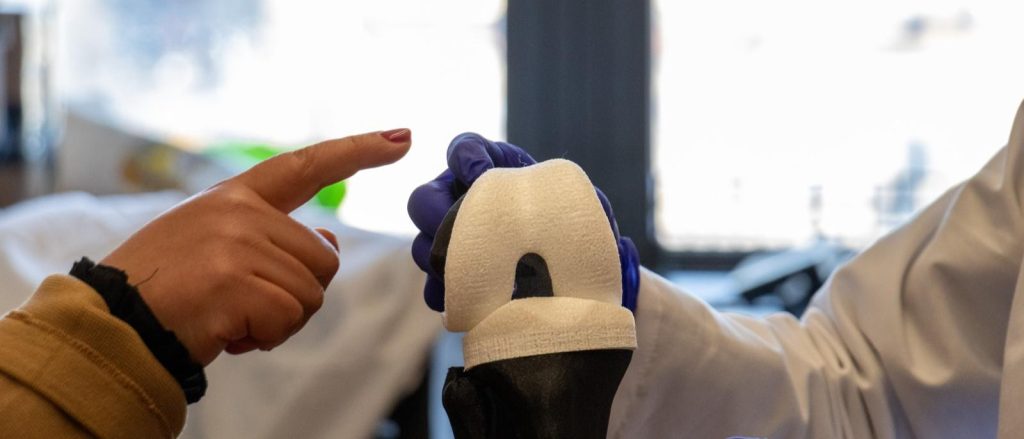
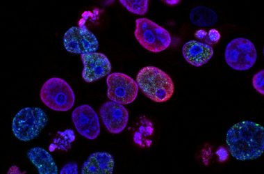
The process begins with medical imaging. A patient’s knee is scanned to produce a precise three-dimensional model of the joint surfaces, both the femoral (thigh bone) and tibial (shin bone) components. This model is then used to 3D-print a polymer scaffold shaped exactly to the patient’s anatomy. The scaffold is seeded with two types of cells: chondrocytes (cartilage cells) on the joint surfaces, and osteoblasts (bone-forming cells) in the deeper structural regions. These cells can be derived from the patient’s own stem cells or from adult induced pluripotent stem cells.
Once implanted, the scaffold serves as a temporary framework. The cells multiply, produce extracellular matrix, and gradually replace the polymer as it degrades. Over time, the synthetic scaffold dissolves entirely, leaving behind a knee joint made of the patient’s own living cartilage and bone. The implant integrates with the surrounding tissue in a way that no metal prosthesis can.
That, at least, is the theory. Preclinical testing, now underway, will determine whether the biology aligns with the engineering. The questions are not trivial. Will the scaffold degrade at the right rate? Too fast, and the joint collapses before the new tissue is strong enough to bear the load. Too slow, and the polymer interferes with tissue ingrowth. Will the cartilage layer maintain its smooth, low-friction surface under the mechanical loads of walking, climbing stairs, and standing from a chair? Healthy knee cartilage withstands compressive stresses of several megapascals thousands of times a day. The engineered replacement must do the same.
Engineering Deep Dive: The science behind the living knee implant
Tissue engineering sits at the intersection of biology, materials science, and mechanical engineering. The NOVAKnee project draws on all three, and A-level students will recognise several underlying principles.
The biodegradable scaffold is made from a polymer, likely a polylactic acid (PLA) or polycaprolactone (PCL) variant, chosen because it degrades through hydrolysis, a chemical reaction with water that breaks the polymer chains into smaller fragments. The body eventually metabolises these fragments into carbon dioxide and water. The degradation rate can be tuned by adjusting the polymer’s molecular weight, crystallinity, and copolymer ratio. A-level Chemistry students studying polymerisation and hydrolysis will find the underlying reactions familiar.
The 3D printing process used to fabricate the scaffold is a form of additive manufacturing, building up the structure layer by layer from a digital model. The printer must achieve sufficient resolution to replicate the complex curvature of the femoral condyles and the tibial plateau, features that vary between patients and are measured in fractions of a millimetre. The scaffold’s porosity is also carefully controlled. Pores must be large enough for cells to migrate into the structure and for blood vessels to grow through it, supplying oxygen and nutrients, but small enough to maintain structural integrity under load.
Stem cell biology is the engine of the regeneration process. Induced pluripotent stem cells (iPSCs) are adult cells that have been reprogrammed back to an embryonic-like state, from which they can be differentiated into almost any cell type. The NOVAKnee team differentiates iPSCs into chondrocytes and osteoblasts using specific growth-factor cocktails, which serve as chemical signals that instruct the cells to adopt particular identities. This is cell signalling in action, a topic covered in A-level Biology under cellular communication and differentiation.
The mechanical engineering challenge is formidable. The human knee transmits forces of up to four times body weight during walking and up to eight times body weight during stair descent. The articular cartilage that lines the joint surfaces is a remarkable material, a hydrated gel of collagen fibres and proteoglycans that distributes compressive loads across the joint while maintaining a coefficient of friction lower than ice on ice. Replicating this combination of load-bearing capacity and lubrication in an engineered tissue is one of the hardest problems in regenerative medicine.
The scaffold must also account for the knee’s complex biomechanics. The knee is not a simple hinge. It combines flexion and extension with rotation and a small amount of translation, guided by the cruciate and collateral ligaments. The NOVAKnee scaffold is shaped to replicate the joint surfaces that interact with these ligaments, preserving the kinematics to which the patient’s own soft tissues are adapted. Getting this geometry wrong would lead to abnormal loading patterns, accelerated wear, and pain.
One particularly elegant aspect of the design is the graded composition of the scaffold. The joint surface layer is optimised for cartilage growth, with a composition and porosity that favour chondrocyte attachment and matrix production. The deeper layers transition to a bone-friendly composition, with larger pores and a stiffer structure that encourages osteoblast colonisation and mineralisation. This gradient mimics the natural osteochondral interface, the transition zone between cartilage and bone that is one of the most mechanically demanding regions in the human body.
If preclinical testing goes well, the first human clinical trials could begin in 2028. NOVAJoint Orthopaedics, a company established to commercialise the technology, is already preparing for regulatory submissions. The road from a promising preclinical result to a routine surgical procedure is long, and there will be failures along the way. But the ambition is clear. Thirty years from now, the idea of bolting a metal hinge into a human knee may seem as crude as wooden teeth.
- Columbia University’s NOVAKnee living knee implant has entered preclinical testing after clearing all ARPA-H milestones
- The implant uses a 3D-printed biodegradable scaffold seeded with stem cells that regenerate cartilage and bone
- The scaffold gradually dissolves as the patient’s own tissue grows to replace it
- The technology could eliminate the need for revision surgeries that current metal implants require after 15-20 years
- If preclinical testing succeeds, the first human clinical trials could begin in 2028