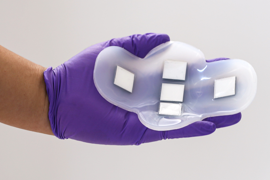
Researchers at MIT have developed a new ultrasound patch that overcomes traditional barriers in ultrasound tech and brings medical imaging out of the lab and into patients’ daily lives.
The flexible, stamp-sized device sticks to the skin for up to 48 hours and provides high-resolution ultrasound images of internal organs without gel or personnel support.

Ultrasound imaging provides a safe, noninvasive, real-time view of the human body’s inner workings. It allows clinicians to visualize a patient’s internal organs and tissues without the risks of radiation exposure. However, conventional ultrasound technology requires bulky machinery, an expert technician to manoeuvre the ultrasound probe, and conductive gel applied to the skin. These limitations have made ultrasound impractical for continuous patient monitoring outside medical facilities.
The tech was partly inspired by a personal experience of bladder dysfunction after kidney surgery. The MIT team wondered if a wearable bladder monitor could help patients like this track their organ health and emptying ability from home. Since bladder volume monitoring already assists clinicians in evaluating kidney issues, the researchers wanted to work on demonstrating bladder imaging with their novel patch.
The ultrasound sticker consists of a silicone rubber adhesive layer embedding an array of tiny ultrasound transducers made from a new piezoelectric material optimized for performance and flexibility. It sticks softly to the skin on its own or can be further secured by clothing. The smooth form factor conforms to the body’s natural contours.
The precise arrangement of transducers within the cross-shaped array enables a wide field of view imaging of the entire bladder region. The elastic polymer casing and hydrated hydrogel layer around the electronics keep the sticker firmly stuck while allowing ultrasonic waves to penetrate deep into tissues. This design ensures precise and accurate visualization of underlying organs throughout body movements.
In tests on human volunteers, the ultrasound patch performed equally well compared to traditional ultrasound probes. It successfully imaged bladder volume changes from empty to complete, and the quality remained reliably high across subjects with diverse body types.
Remarkably, the stickers required no manual application of pressure or gel. The inherent adhesion kept transducers coupled to the skin, while the large field of view covered the bladder without repositioning. These features enable the possibility of continuous ultrasound monitoring without a human technician.
This novel tech holds promise far beyond bladder monitoring. By tuning technical parameters like transducer frequency and location on the body, the patch can image many organs for various diagnostic and monitoring applications.
The team aim to design customized patches for imaging various parts of the abdomen, like the liver, kidneys, and ovaries. Smaller configurations could also fit on the neck or chest. The stickers would provide vital clinical information without invasive procedures or compression garments.
In the future, patch technology may enable the early detection of tumours and cancers. Integrating wireless communication and AI analysis could also make wearable ultrasound practical for rapid triage and telemedicine. This disruptive advancement could take ultrasound imaging out of dedicated facilities and into patients’ daily lives, providing clinicians with richer insights into disease progression and health while improving diagnostics and access to care.
Read the paper at nature.com
TLDR:
- MIT developed a revolutionary ultrasound sticker that can image organs continuously without gel.
- The flexible, stamp-sized patch contains a specialized ultrasound array optimized for wearability.
- In initial tests, it accurately monitored bladder volume changes when worn on human volunteers.
- It may allow ultrasound imaging of many organs for improved diagnostics and monitoring.
- The technology could enable early cancer detection and take ultrasound outside of labs into daily life.


