
Spinal Cord Stimulation Restores Walking and Sensation in Paralysis Trial
A clinical trial shows spinal cord stimulation can restore walking and sensory feedback in people with complete spinal injuries, a biomedical engineering first.
Key points
- Three volunteers with complete spinal cord injuries walked on a treadmill using implanted electrode arrays that deliver patterned electrical stimulation
- For the first time, stimulation was applied both above and below the injury site, restoring motor control and sensory feedback simultaneously
- Machine learning algorithms optimised stimulation patterns across an enormous range of possible settings
- No adverse effects were reported, clearing the way for longer-term clinical studies outside hospital settings
When a spinal cord is completely severed, two things vanish at once. The brain can no longer tell the legs to move, and the legs can no longer tell the brain where they are. That second loss, the disappearance of sensation, is the one that most people outside medicine rarely think about. You cannot walk if you cannot feel the floor.
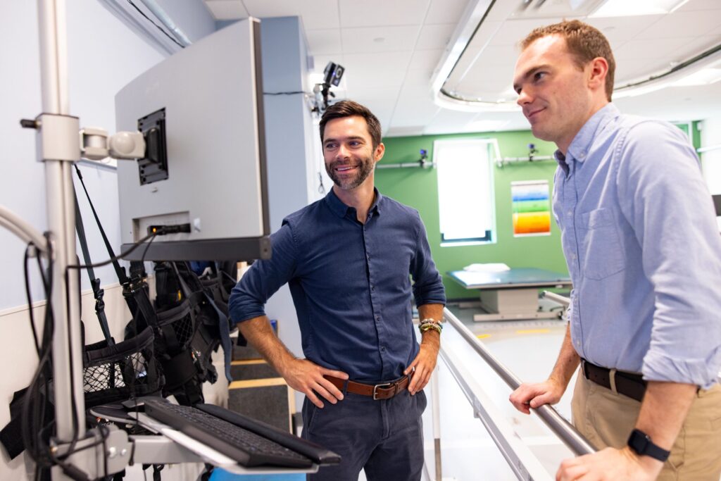
A clinical trial at Brown University has now demonstrated, for the first time, that both problems can be tackled simultaneously using electrical stimulation. Three volunteers with complete spinal cord injuries walked on a treadmill while implanted electrodes delivered carefully patterned signals above and below their injury sites. Electrodes below the lesion restored enough muscle control for stepping. Electrodes above it produced sensations, felt in the chest, back, or arm, that the participants learned to interpret as information about where their legs were in space. The results, published by Brown University in March 2026, represent a convergence of biomedical engineering, neuroscience, and machine learning that has been decades in the making.
Two arrays, one problem, and a DJ board
The engineering setup is more inventive than it first appears. Surgeons implanted small electrode arrays at two locations on each participant’s spinal cord: one above the injury and one below. The sub-lesion array targeted motor neurons in the lumbar region, the stretch of spinal cord that controls leg movement. By delivering patterned electrical pulses, it activated the neural circuits responsible for flexing and extending the leg, the basic rhythm of a walking stride.
The supra-lesion array, placed above the injury, tackled the sensory problem. Because the damaged cord blocks sensory signals from travelling up to the brain, the researchers used a technique they call sensory replacement. Electrical stimulation above the break produced sensations that participants could feel, such as tingling or pressure in the chest. The intensity of those sensations was linked to the angle of the participant’s knee joint, measured by external sensors. With practice, participants learned to associate a particular chest sensation with a particular leg position. It is not natural proprioception. It is an engineered substitute, and it worked well enough for participants to report accurately where their knees were during walking.
The control interface for the stimulation is a device the research team nicknamed the “DJ board.” It has knobs and sliders that adjust stimulation patterns, intensity, and electrode selection. David Borton, an associate professor of engineering at Brown and the study’s senior author, has spent years refining how electrical parameters map onto biological outcomes. The challenge is scale: each electrode array has multiple contacts, each capable of delivering different pulse widths, amplitudes, and frequencies. The number of possible stimulation combinations is enormous.
That is where machine learning enters the story. An algorithm developed by Thomas Serre’s laboratory at Brown, built by researcher Lakshmi Narasimhan Govindarajan, analysed data from the DJ board sessions and identified which stimulation settings produced the best muscle activation. Rather than testing every combination manually, the algorithm learned to predict effective settings, dramatically reducing the time needed to calibrate each participant’s system.
Engineering deep dive
How spinal cord stimulation restores walking
The spinal cord is not simply a cable that carries instructions between the brain and the body. It contains its own processing circuitry, including networks of neurons called central pattern generators (CPGs) that can produce rhythmic motor patterns, such as the alternating flexion and extension of walking, without direct input from the brain. This is why a decerebrate cat, one with the connection between brain and spinal cord severed, can still produce stepping movements on a treadmill. The motor circuitry for walking lives in the cord itself.
Epidural electrical stimulation exploits this architecture. By placing electrodes on the surface of the spinal cord in the lumbar region (typically around vertebrae L1 to S1) and delivering tonic or patterned pulses, engineers can raise the excitability of the CPG circuits to a threshold where they begin firing in coordinated patterns. The stimulation does not directly command individual muscles. Instead, it sets the conditions for the cord's own circuits to generate movement. Think of it as turning up the volume on a signal that was too quiet to hear.
The physics of the electrode-tissue interface is governed by charge injection. Each electrode delivers a biphasic current pulse, typically with a leading cathodic phase that depolarises nearby neurons. The charge density at the electrode surface must stay within safe limits, roughly 30 microcoulombs per square centimetre, to avoid tissue damage. Electrode materials such as platinum-iridium are chosen for their electrochemical stability and high charge injection capacity. Students studying A-level Physics will recognise the principles here: current, charge, and potential difference, applied at the boundary between electronics and biology.
The sensory replacement system is an engineering workaround for a biological barrier. Normally, proprioceptive signals travel from muscle spindles and joint receptors through sensory neurons in the dorsal root ganglia, up the dorsal columns of the spinal cord, and into the brain. A complete injury blocks this pathway entirely. The Brown team's approach bypasses the block by generating artificial sensations above the injury that encode the same information. The brain does not care where a signal comes from, only what it represents. With training, participants re-map the new signals onto their existing body schema.
The machine learning component addresses a practical bottleneck. With multiple electrode contacts, each accepting a range of amplitudes (0 to 10 milliamps), pulse widths (50 to 500 microseconds), and frequencies (2 to 120 Hz), the parameter space for a single array runs into millions of combinations. Exhaustive search is impossible. The optimisation algorithm uses a form of Bayesian inference, maintaining a probabilistic model of which parameter regions produce good outcomes and focusing its search there. This is closely related to techniques used in A-level Further Maths, where iterative methods converge on optimal solutions without evaluating every possibility.
The safety data from this trial is as significant as the functional results. No device-related adverse effects were reported across all three participants, which is the minimum threshold for progressing to larger, longer-term studies. The electrode arrays remained stable and functional throughout the trial period, suggesting that the materials and surgical placement protocols are robust enough for chronic implantation.
What separates this work from earlier spinal stimulation research is the simultaneous restoration of motor and sensory function in the same participant. Previous studies have demonstrated that sub-lesion stimulation can elicit stepping. Others showed that cortical or peripheral interfaces could provide feedback. Putting both together in a single, coordinated system is the engineering advance that makes functional walking, rather than reflexive leg movement, possible.
Links to the curriculum: A level, GCSE
The team plans to recruit new participants for a longer-term study that will test stimulation outside the hospital, in homes and rehabilitation centres. Whether this technology can scale from a three-person trial to a clinical product depends on manufacturing the electrode arrays reliably, making the machine-learning calibration fast enough for routine clinical use, and securing funding for the regulatory pathway. The gap between a treadmill in a laboratory and independent mobility in the real world remains wide. But for three people who had been told they would never feel the ground again, that gap just narrowed.
Credit via brown.edu
Share
Related stories
All Health
Electronics & CommunicationsBreakthroughs
Open Bionics Pioneers 3D Printed “Hero Gauntlet” for Partial Hand Amputees
Electronics & CommunicationsBreakthroughs
Printed Artificial Neurons Communicate With Brain Cells

Electronics & CommunicationsBreakthroughs